Nanoparticle Synthesis Laboratory (BIOMA)
Synthesis
Description
Nanoparticle Synthesis Laboratory - Bionanoplasmonics laboratory (BIOMA)
The relevance of noble metal nanostructures is due to their physical and chemical properties, which can be tailored not only by varying material composition, but also by tuning the size and shape of the nanostructure. Control over composition, size and morphology at the nanoscale is of high current interest due to the remarkable effects that small changes produce on a variety of physical (optical, magnetic, catalytic, etc.) properties of a material.
The Bionanoplasmonics Laboratory at CIC biomaGUNE (BIOMA) has developed synthetic strategies toward creating a toolbox of structurally uniform noble metal nanoparticles in high-yield, with excellent control over their shape and size. We exploit colloid chemistry as a tool to prepare a wide variety of nanoparticles with tight size and shape control. We are thus interested in understanding nucleation and growth processes, which are crucial steps during the synthesis of a nanocrystal. In particular, we focus on so-called seed-mediated syntheses, which are based on using metal seeds as templates for the growth of the desired nanoparticle products. By separating the processes of (seed) nucleation and (nanoparticle) growth we can better control the mechanisms behind seed-mediated synthesis, enabling a high degree of control over the structural uniformity and yield of the nanostructures. The ultimate goal of research developed at BIOMA is to build a scientific base through fundamental understanding of the processes for the production of nanocrystals with specific properties for applications in electronics, photonics, catalysis, optical sensing, and biomedical research.
We have additionally developed strategies to fabricate a variety of hybrid nanomaterials with different configurations (single-component, core-shell, alloys): multimetallic systems comprising (Ag, Pd, Pt, Cu), oxides (silica, mesoporous silica, TiO2), polymers (polyethyleneglycol, polystyrene, poly-NIPAM).
Access is provided to a state of the art synthesis infrastructure, as well as to technical and scientific support by local engineers and scientists, thereby ensuring adequate training, experimental support and supervision. The broad range of techniques made available through BIOMA institutional facilities enable a complete characterization of the nanomaterials whenever necessary (UV-Vis-NIR, TEM, SEM, ICP-MS, Z-potential, light scattering, AFM, NMR, IR, Raman, XPS, etc.). Training of characterization techniques may also be provided.
Services currently offered by the infrastructure
External users have access to a wide variety of nanomaterials that are currently available at the Bionanoplasmonics Laboratory, with adequate training and supervision by the BIOMA scientists who have developed the relevant procedures to synthesize the nanomateriales. The EUSMI projects are performed within our laboratories, so that the environment is amenable to frequent discussion. In addition, users have access to a fully equipped synthesis facility at BIOMA, the Nanofabrication Platform, which is led by a researcher with a wide expertise in nanomaterials synthesis, self-assembly and surface modification.
Nanomaterials currently offered by the infrastructure
Spherical gold nanoparticles

Figure 1. TEM images and UV-Vis spectra of monodisperse gold spheres with different diameters.
Anisotropic gold nanoparticles
Gold nanostars
 Figure 2. TEM images and UV-Vis spectra of PVP-coated gold nanostars of different sizes.
 Figure 3. TEM images and UV-Vis spectra of citrate-functionalized gold nanostars.
Gold nanorods
 Figure 4. TEM images and UV-Vis spectra of single-crystalline gold nanorods with different aspect ratios.
Other anisotropic gold nanoparticles

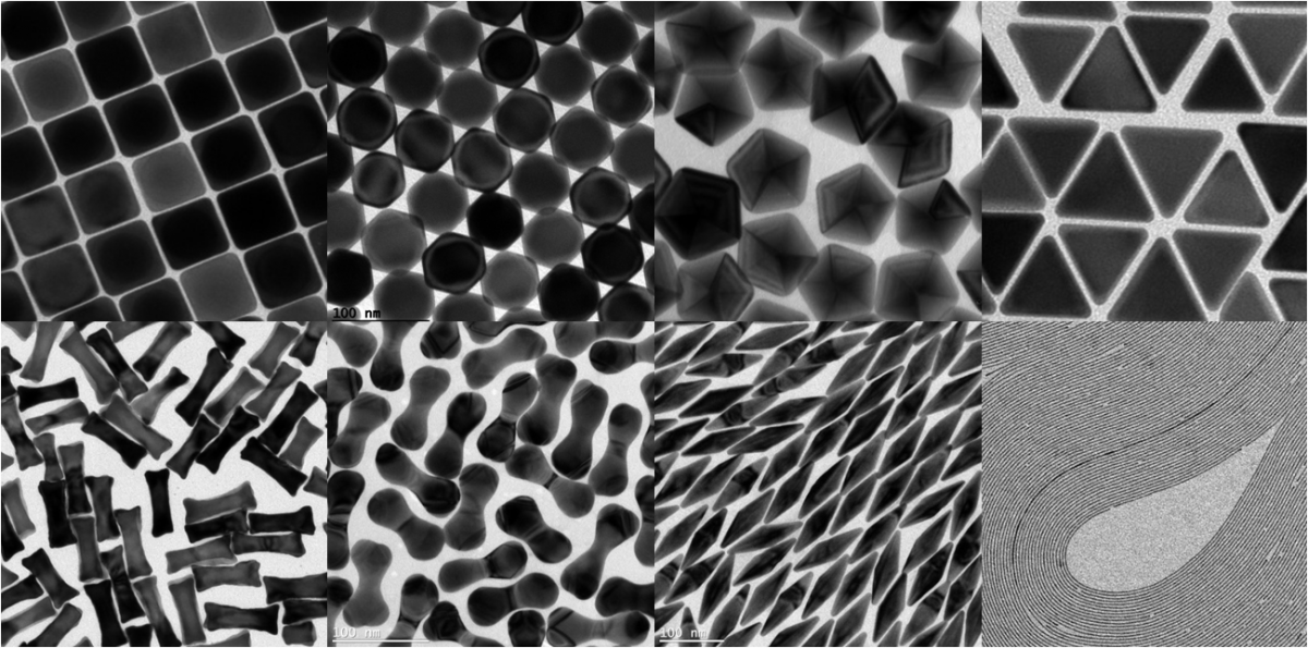
Figure 5. TEM images of gold nanoparticles with different shapes: cubes, octahedra, decahedra, triangular plates, dog-bones, dumbbells, bipyramids and wires.
Silica-coated gold nanoparticles
Control over silica shell thickness Figure 5. TEM images and UV-Vis spectra of spherical gold nanoparticles coated with silica shells of varying thicknesses.
Gold nanoparticles coated with mesoporous silica shells

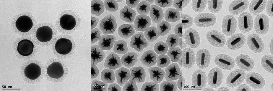
Figure 6. TEM images of gold nanoparticles with different shapes (spheres, stars and rods), coated with mesoporous silica.
Silver-coated gold nanoparticles
 Figure 7. TEM images and UV-Vis spectra of gold nanorods (black line) and silver coated gold nanorods (red line).
 Figure 8. TEM images and UV-Vis spectra of gold bipyramids (black line) and silver coated gold bipyramids (red line).
Equipment currently offered by the infrastructure
BIOMA is equipped with state-of-the art techniques available for users upon request, including:
Transmission and Scanning Electron Microscopy:
- JEOL JEM-2100F UHR (80kV - 200 kV)
- JEOL JEM-1400PLUS (40kV - 120kV)
- JEOL JSM-6490LV
UV-Vis-NIR and Fluorescence spectroscopy:
- Varian Cary 5000 UV-Vis-NIR spectrophotometer
- Agilent UV-Vis spectrophotometer 8453
- Perkin Elmer LS55 fluorometer
Size and Zeta potential characterization:
- Zetasizer Nano ZS
ICP-MS:
- Thermo ICP-MS iCAP-Q
Raman scattering:
- Renishaw Raman Confocal Microscope
XPS characterization:
- SPECS SAGE HR 100
Local Contacts
- Prof. Luis M. Liz-Marzán
- Miss Ana Sánchez Iglesias
